
Diabetic Eye Problems
Eye problems are a significant complication of diabetes and are the most common cause of blindness in people of working age.
Diabetes can affect the eyes in a number of ways;
- If undetected and untreated it can cause noticeable changes to your eye sight.
- If poorly controlled, it can damage the fine network of blood vessels at the back of your eye and cause them to leak fluid.
- Diabetes increases the occurrence of cataracts.
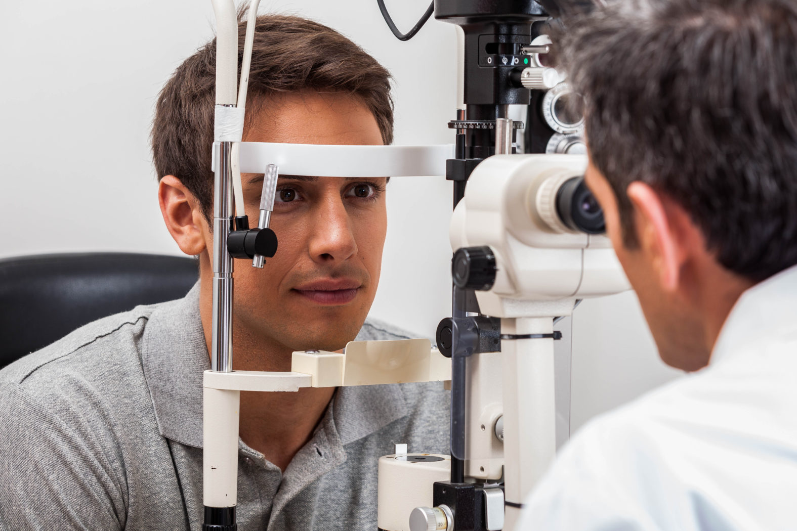
Optometrists play a role in detecting and monitoring diabetic eye disease. Drops to aid pupil widening are used and a careful examination of the back of the eye (retina) is conducted. Retinal photos are invaluable in helping to detect and monitor any changes. If any serious diabetic eye disease is found, referral to an ophthalmologist is arranged for treatment. However, most sight loss from diabetic eye disease can be prevented if detected early and treated.
If you are diabetic you are entitled to an annual NHS funded eye examination.
For more information please click on the following links;
Glaucoma
Glaucoma is defined as a group of conditions where there is progressive damage to the optic nerve. This results in a gradual loss of peripheral field of vision. Raised pressure within the eye is a risk factor towards developing glaucoma. Other risk factors include:
- Increasing age. After the age of 40 risk of glaucoma increases with each decade. By the age of 80 the risk increases to 1 in 10 of us developing glaucoma .
- Family history particularly a first degree relative such as parent, child, sibling.
- Certain ethnic groups
- Diabetics
- Those who are very short sighted
Early detection is vital to the long term prognosis of this condition. Most cases of glaucoma are identified through referrals from Optometrists. The 3 main tests that an Optometrist will perform to check for glaucoma include;
- Ophthalmoscopy – to examine the appearance of the optic nerve
- Visual Field Test – to check for any blind spots in your field of vision.
- Tonometry – to measure the pressure of the fluid within your eyes.
As most types of glaucoma are not symptomatic until advanced, regular eye examinations are important in screening for this condition. Any suspicious signs will be referred to an ophthalmologist.
NHS funded eye examinations are available if;
- You are over 40 and have a parent, child, sibling with glaucoma
- You have glaucoma or are considered to be at risk of glaucoma by an ophthalmologist.
For more information please click on the following links;
RNIB
International Glaucoma Association
Cataracts
A cataract is where the transparent, internal focusing lens of the eye starts to become cloudy. This restricts the amount of light that can pass through to the back of the eye. Visual symptoms include:
- Blurred, hazy vision
- Glare in bright light
- Colours appear faded
- Shadowy/doubled vision
Cataracts occur naturally with increased age and for most people they progress slowly. They can also be caused by head trauma, diabetes and certain medication.
In the early stages no treatment is necessary. Changes in the spectacle prescription and sunglasses will help to enhance vision. As the cataract progresses the only solution is to have surgery.
Cataract surgery is usually done as day-case under local anaesthetic. It involves removal of the cloudy lens and replacement with a clear artificial lens. The results are instantaneous and people often comment on how sharp and bright their vision feels.
For more information please click on the following links:
RNIB
Macular Degeneration (AMD)
AMD is the most common reason for becoming registered visually impaired in the western world. Some statistics suggest 1 in 10 people over 55 are affected by this condition.
The macula is the central area of the retina and its function is to provide fine detailed central vision. In AMD this function is disrupted resulting in distorted and degraded central vision. Tasks such as reading, writing, recognising facial features become increasingly difficult.
There are 2 main types of AMD;
DRY – this is more common and has a more gradual onset. Although there is no treatment to reverse the damage, help in the form of counselling, nutritional advice, magnifiers and other practical aids will improve life quality.
WET – this form is less common, much more severe and has a more sudden onset. This requires urgent referral to an ophthalmologist for treatment.
AMD is primarily an age related condition but there are other risk factors;
- smoking
- a diet high in saturated fat
- high blood pressure and high cholesterol
- UV exposure.
Making the right lifestyle changes from a young age can significantly reduce the risk of developing AMD. These changes include:
- Stop smoking
- Protect your eyes from UV light with regular use of quality sunglasses
- Eat a balanced diet with green leafy vegetables like spinach and kale, omega 3 rich foods such as nuts, fish and eggs.
- Get regular exercise.
Nutritional supplements specific for AMD are available. A new product called Macushield specifically contains all 3 macular pigments. Research shows that this supplement enhances macular pigment levels in those with dry AMD and in some sufferers has helped to significantly improve vision over a period of time. Increasing the macular pigment levels gives this delicate area its protective shield back against oxidative stress and UV damage which is thought to cause AMD. The contents of Macushield are derived from the marigold flower and this supplement does not conflict with other medication.
Regular eye examinations aided by enhanced macular examination using dilating drops and retinal photography will allow earlier detection and treatment.
Please click on the flowing links for more info;
RNIB
Macular disease society
Strabismus / Amblyopia
An eye examination will also screen for amblyopia (lazy eye) and strabismus (turning eye/squint)
Amblyopia = where one eye is weaker and cannot see the same amount of detail as the fellow eye. This can prevent the two eyes working together and make depth perception and binocular vision difficult. Amblyopia tends to occur when only one eye is significantly long-sighted, short-sighted or astigmatic and corrective glasses are not prescribed early enough. Also if one eye has a strabismus (squint) it may also become amblyopic. The main method of treatment is patching of the good eye for several hours a day in order to stimulate the weak eye. Rarely there may be an organic reason such as a cataract or retinal disease for amblyopia.
Strabismus = where one eye appears to turn in, out, up or down, either constantly or when concentrating or tired. This will also disrupt development of normal binocular vision. Children do not outgrow squints and appropriate treatment and monitoring at the earliest possible stage is vital.
Both these conditions are prevalent in childhood and have many different causes. They need to be diagnosed and investigated promptly. Early intervention will allow a chance for normal sight to develop. If necessary your child can be referred to a paediatric ophthalmologist for further treatment.